The diaphragm muscle divides the thoracic and abdominal cavities and serves as the principal respiratory muscle.
Active downward movement or contraction of the diaphragm generates an increase in negative pleural pressure that causes the lungs to expand and fill with air during inspiration.
Passive upward movement or relaxation of this crucial muscle causes an increase in pleural pressure and results in an increased pressure in the airways, facilitating expiration. The diaphragm is innervated by the phrenic nerve , which originates from the C3, C4, and C5 spinal nerves. In addition to the motor fibers to the diaphragm, the phrenic nerve also contains pain fibers, which is why pain originating from the diaphragm can be referred to the shoulders.
Attachments
The diaphragm is a musculotendinous structure that separates the thoracic cavity from the abdominal cavity. The diaphragm is composed of a peripheral muscular portion and a central tendon (Figure A).
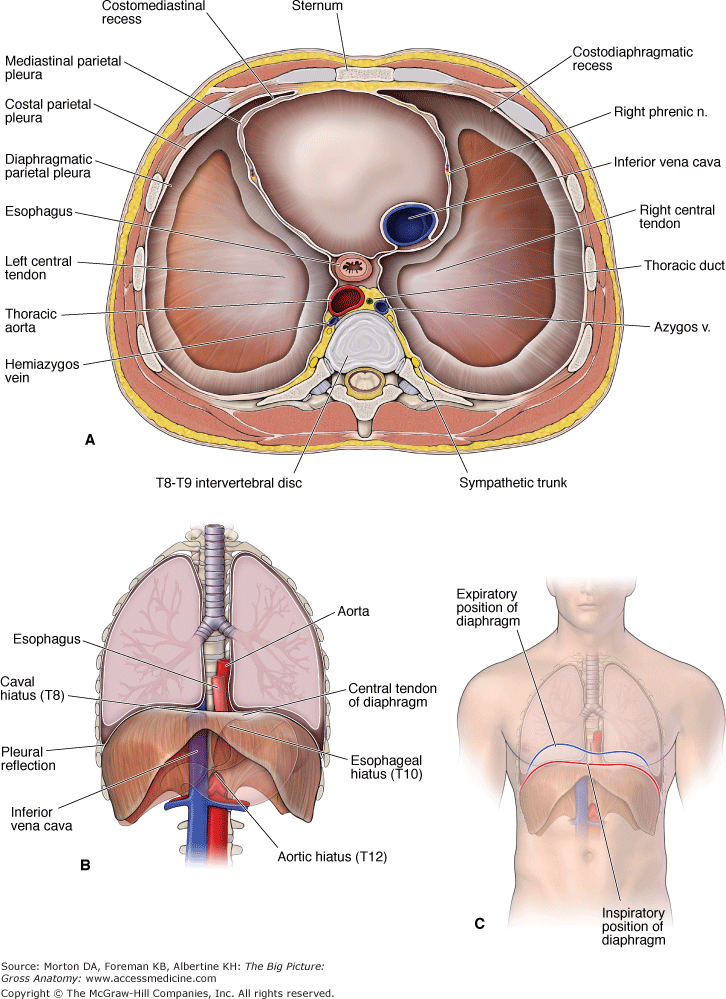
A. Superior view of the axial section of the thorax above the diaphragm. B. Anterior view of the diaphragm and its relationship to the lungs. C. Position of the diaphragm during inspiration and expiration.
The diaphragm is dome shaped, and upon contraction of its muscular portion, it descends (flattens). The right dome, resting atop the liver, is generally higher than the left dome, which rests atop the fundus of the stomach.
The muscular portion has three regions of origin (Figure B):
- Lumbar origin. Two crura originate from the bodies of the upper two (left crus) or three (right crus) lumbar vertebrae.
- Costal origin. From muscle fibers that arise from the inner surfaces of the lower six ribs.
- Sternal origin. From muscle fibers that arise from the inner surface of the xiphoid process.
The muscle fibers of the diaphragm extend centrally to insert into its central tendon. The central tendon is the structure that the diaphragm's muscle fibers primarily pull upon when they concentrically contract.
Innervation
The right and left phrenic nerves provide sensory and motor innervation to the diaphragm as follows:
- Sensory. Sensory innervation of the pericardium, mediastinal and diaphragmatic pleurae, and the diaphragmatic peritoneum.
- Motor. Motor innervation of the diaphragm is through the phrenic nerve, which arises from the cervical plexus via ventral rami branches of C3, C4, and C5 (“C3, C4, and C5 keep the diaphragm alive”).
The phrenic nerves course along the anterior surface of the anterior scalene muscle in the side and descend into the thoracic cavity between the subclavian vein and artery. The phrenic nerves are accompanied by the pericardiacophrenic vessels and descend anterior to the root of each lung between the mediastinal pleura and pericardium en route to the diaphragm. The vagus nerve, by contrast, courses posterior to the root of the lung.
Apertures in the Diaphragm
The diaphragm has several apertures that permit the passage of structures between the thorax and the abdomen (Figure A and B). The caval, esophageal, and aortic openings are large, and the left and right crura provide other small openings.
- Caval opening. Located at the T8 vertebral level within the central tendon of the diaphragm, just right of the midline. The caval opening allows passage for the inferior vena cava and branches of the right phrenic nerve. Branches of the left phrenic nerve pass through the diaphragm by piercing the central tendon on the left side.
- Esophageal opening. Located to the left of the midline at the T10 vertebral level. The opening usually splits the muscle fibers of the right crus. The esophageal opening allows passage for the esophagus and the left and right vagus nerves. The esophageal branches of the left gastric artery and the esophageal tributaries of the left gastric vein also passes through this opening.
- Aortic opening. Located at the T12 vertebral level, behind the two crura. Strictly speaking, the aortic opening is not an opening through the diaphragm but rather a large gap between the crura. The left and right crura form the left and right borders of the aortic opening. The aortic opening allows passage for the aorta, the azygos vein, and the thoracic lymphatic duct.
- Right and left crura. The right and left greater and lesser splanchnic nerves course from the thoracic cavity, deep to the right and left crura en route to the prevertebral ganglia of the abdomen. In addition, the left crus also allows passage for the hemiazygos vein.
Functions of the Diaphragm
- Respiration. The diaphragm is the principal muscle of inspiration. It flattens upon contraction, thus increasing the vertical dimensions of the thoracic cavity (Figure C). The roles of the diaphragm and other thoracic muscles of respiration are discussed in more detail in Chapter 3.
- Venous return. The alternating contraction and relaxation of the diaphragm causes pressure changes in the thoracic and abdominopelvic cavity that facilitate the return of venous blood to the heart.
 Valsalva maneuver. When taking and holding a deep breath, an individual forcibly contracts the diaphragm inferiorly on the abdominal viscera, thereby increasing the pressure in the abdominal cavity. This is done to help expel vomit, feces, and urine from the body by increasing the intra-abdominal pressure and by preventing gastric reflux by exerting pressure on the esophagus as it passes through the esophageal hiatus.